Decoding diseasein the bone marrow
Redefine blood cancer insights with our AI-driven platform
Quantify established markers, uncover novel morphological patterns predictive of outcomes, and contextualize findings within our curated patient cohort.
Built at the world's leading research organization ↘︎


Modular markers
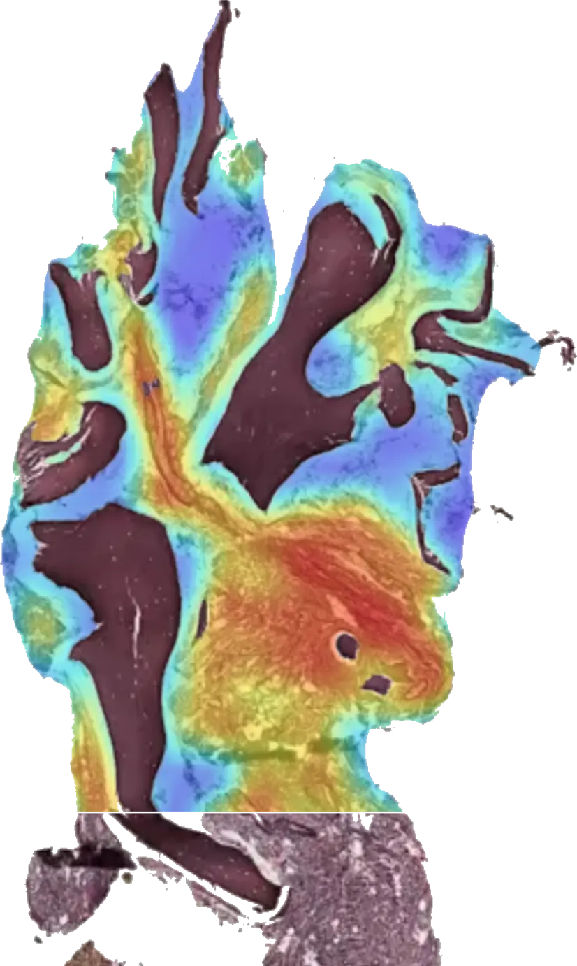
An integrated suite of AI models for quantifying morphology in bone marrow
Measure key features in bone marrow tissue—such as fibrosis, megakaryocytes, blasts, and plasma cells—at both the slide and cell levels. Leverage our platform to streamline analysis, enhance reproducibility, and unlock new insights for clinical and research applications.

Novel prognostics
Predict outcome directly from morphology
Use our AI-driven discovery platform to uncover novel morphological patterns in bone marrow tissue that predict clinical outcomes, such as disease transformation or adverse events. We're working towards new actionable insights to inform patient care and guide therapeutic decision-making.
Cohort-wide context
Compare results across diverse patient cohorts
Place findings in the context of your whole cohort - and then map it against our diverse disease-defining datasets. Gain a deeper understanding of disease variability and refine insights with robust cross-cohort comparisons.

All from standard-of-care slides
Get more from your existing data and workflows — no special staining or obscure data requirements.
Boost your research with the AI-driven platform for bone marrow morphology
We're applying our technology to trials, research, and clinical audits today.
Get in touch